Compared to using CT scans, planning, determining and practicing optimal surgical approaches with 3D printed models can enable better understanding and treatment of complex cases. “On average, 1,000 to 2,000 images are made per CT scan in vascular-related patient cases, which surgeons use to analyze and diagnose the illness. This can be ambiguous and time-consuming when the issue is complex,” said Dr. Dorweiler. “With 3D printed models, we can quickly understand the cause of the issue and determine the best type of treatment.”
Recently, a patient turned down by several hospitals because of the complexity of her illness was treated at the University Hospital Mainz. Due to an aortic malformation close to her heart, the patient was suffering from a bulging blood vessel on her neck. “Looking at the CT scans, it was impossible to clearly visualize the anatomy,” said Dr. Dorweiler. “So we 3D printed a model, and for the first time it became clear what the origin and magnitude of the problem was. Not only did we use the model to explain our findings to the patient, we took it into all three surgeries as a point of reference.”
Treating complex aortic illnesses with the endovascular method, the affected blood vessel is replaced and supported by an endovascular stent, a type of implant in the form of a small wire-mesh tube. The stent is inserted through the arteries and placed at the affected area of the aorta. This very precise and difficult procedure means the surgeon is operating using a monitor, and has no room for error. As such, the stent itself needs to be perfectly designed to fit the patient’s anatomy to mitigate risk.
“By 3D printing a model of the patients’ blood vessel where the stent needs to be placed, we save significant time and money as we can practice surgery on the model repeatedly until we are certain everything fits together and that we have perfected the procedure,” said Dr. Dorweiler. In planning for another very complex case of aortic arch aneurysm, the team underwent a pre-operative simulation of the surgery using a stent prototype and a 3D printed aortic arch model of the patient. This helped ensure the correct design and fit of the stent, and helped the team understand exactly where it should be placed, significantly reducing operating time.
“Based on current studies, we are seeing savings in operating time of 5-45 minutes when using 3D printed models prior to surgery,” said Dr. Dorweiler. “Research is still ongoing, but if you take an average surgery time of two to four hours, you are looking at time savings of up to 40%. When you are dealing with complex vascular cases every day, these time-savings can be the difference between life and death.”
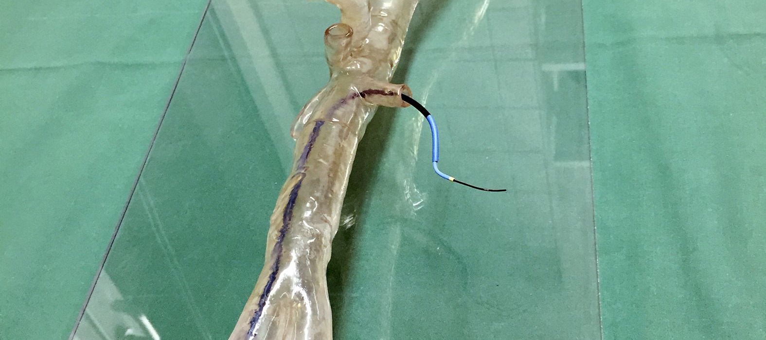
The University Hospital Mainz also has an extensive research and training facility where 3D printing is used to give future surgeons hands-on, realistic training. “We use the Stratasys Eden260VS 3D Printer in our BiomaTiCS research platform to produce models of aortic anatomies from real-life cases to teach future vascular surgeons how to successfully perform complex surgeries,” said Dr. Dorweiler. “With the ability to 3D print ultra-realistic aortic models, the trainees can practice endovascular procedures and learn difficult wire-skills using the accurate replicas of blood vessels. For healthcare, it is crucial that we continue to leverage the capabilities of 3D printing for medical training, education and research for future breakthrough-implementation.”

